
No products in the cart.
What is Skin Cancer?
Overview
Skin cancer is that the most typical kind of cancer, in all probability creating up over half all diagnosed cases of cancer, consistent with the yankee Cancer Society (ACS). The incidence of carcinoma is rising dramatically within the us. More than three million cases of non-melanomaare diagnosed each year, leading to about 3,000 deaths. And the ACS estimates that in 2012, there will be 76,250 new cases of melanoma and 9,180 deaths from the disease. In fact, between forty p.c and fifty p.c of individuals within the us over age sixty five can develop non-melanoma carcinoma. This type of cancer is highly treatable when diagnosed in its early stages and is usually relatively easy to diagnose.
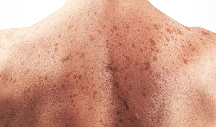
The majority of life sun exposure happens before age twenty, and skin cancer can take 20 years or more to develop. In fact, very young children who experience as few as two to three severe sunburns are believed to have an increased risk of developing skin cancer later in life. That’s to not say you must ignore your risk of developing carcinoma. You need to be concerned about skin cancer, whether your sunbathing days are over or you still spend time pursuing the perfect tan.
The Structure of Skin
The skin is the largest organ in your body and is the body’s first defense against disease and infection. It also protects your internal organs from injuries. The skin regulates temperature, prevents excess fluid loss and helps to remove excess water and salt from your body.
Skin is composed of two layers: the epidermis (the outermost layer of skin) and the dermis (the lower layer). The stratum itself has four layers: the stratum, the granular layer, the epithelial cell layer and therefore the basal cell layer. Keratin (dead, dense protein cells) makes up the stratum corneum or outer layer of the epidermis—the skin layer that can be seen and felt.
The granular layer moves the dead albuminoid cells to the surface of the stratum. The epithelial cell layer produces albuminoid for the stratum and conjointly transports water. The basal cell layer is that the lowest layer of the stratum. This is wheresquamous cells are produced and where the cells that produce melanin, or skin pigment, reside.
The dermis is the deeper layer of skin. It is a diverse combination of blood vessels, hair follicles and sebaceous glands or oil glands. The proteins collagen and elastinare found in the dermis. They provide support and elasticity to the skin. The sun’s rays eventually break down these proteins. With age, the skin naturally begins to wrinkle and sag.
The body covering level, or subcutis, is a layer of fatty tissue that provides nourishment to the dermis and upper layers of skin. It conjointly conserves body heat and cushions internal organs against trauma. Blood vessels, nerves, sweat glands and deeper hair follicles are found here.
Types of Skin Cancer
There area unit 2 main teams of carcinoma: non-melanoma skin cancer, the most common type of skin cancer, and melanoma (sometimes referred to as “malignantmelanoma”) skin cancer.
According to the ACS, basal cell carcinoma makes up 80 percent of non-melanoma skin cancers, and squamous cell carcinomas account for about 20 percent. Together, these two types account for about 95 percent of all new cases of skin cancer. Over three million cases of non-melanoma skin cancer are diagnosed every year in the United States. Men have a better risk than girls of developing these skin cancers.
Melanoma is that the least common, but most aggressive, of the three types of skin cancer. It originates within the skin’s melanocytes—the cells that turn out pigment, or melanin.
In 2012, the ACS estimates that 76,250 new cases of melanoma will be diagnosed in the United States—about 4 percent of all diagnosed skin cancers. But melanoma accounts for about 75 percent of skin cancer deaths. One person dies of skin cancer virtually each hour (every sixty two minutes).
Risk Factors
Anyone can develop skin cancer, but people with fair complexions are more susceptible to precancerous conditions and skin cancer than people with darker skin tones. That’s because darker skin has more melanin, which provides some natural protection against the sun’s damaging rays. In addition to fair skin, other risk factors for skin cancer include:
- a personal history of skin cancer
- a tendency to freckle or burn easily
- lots of sun exposure throughout your life
- many sunburns as a child or adolescent
- family history of the skin cancer or conditions that are more likely to develop into skin cancer
- chronic, non-healing scarring
- radiation therapy
- exposure to toxic materials, such as arsenic, coal tar or creosote
- exposure to certain subtypes of human papillomavirus (genital warts) especially in people with compromised immune systems
- taking immunosuppressant drugs (after an organ transplant, for instance)
Diagnosis
Health care professionals are able to evaluate many skin abnormalities. A medical aid medico ought to be the primary health care skilled you see if you notice one thing suspicious on your skin. Then you may ask a skin doctor, a physician with extensive training in skin care and skin disorders, particularly skin cancer.
The first step in detection abnormalities which will be carcinoma begins with you. Examine your skin once a month for any suspicious changes. Look for changes in color, size and surface texture of a mole. Sores that will not heal may additionally indicate cancerous or malignant neoplasm conditions of the skin that require attention.
Actinic keratoses. This precancerous condition typically occurs in people with a long history of sun-damaged skin. Lesions appear as rough, crusty bumps on the back of the hands, lips, face, scalp or neck that may itch or feel tender on sun-exposed skin. They may be pink or white. If untreated, these bumps may develop into skin cancer. They affect more than 10 million Americans and are usually more prevalent in people over 40 and in sunnier climates. However, they may show up earlier in people who have used tanning beds or sun lamps.
Basal cell carcinoma. Basal cell carcinoma show up as flat, firm, pale areas or as small raised pink or red pearly bumps that may bleed after minor injury. These bumps or growths may appear anywhere on the body regularly exposed to the sun, such as the head and neck. They are slow growing and rarely spread to other parts of the body. But they’ll extend deep into the skin, causing significant local damage. Approximately 80 percent of all skin cancer cases diagnosed annually are basal cell carcinoma. This form of carcinoma features a high cure rate. However, if left untreated, basal cell carcinoma can result in disfigurement.
Squamous cell carcinoma. The second most common non-melanoma skin cancer, squamous cell carcinoma, appears as nodules or as red, scaly patches, typically on the ears, the face, the lips and mouth. These patches eventually develop into large masses. According to the Skin Cancer Foundation, more than 700,000 cases of this type of cancer are diagnosed each year, leading to about 2,500 deaths. This type of skin cancer is slightly more likely than basal cell carcinoma to spread to other parts of the body. But it is also highly treatable.
Melanoma: willcer} can develop from a pre-existent mole or on clear, sleek skin. Unlike a noncancerous mole, melanoma is irregularly shaped or has irregular borders, and is black, brown or tan. Melanoma is rare in childhood and adolescence, but it is one of the more common cancers in younger adulthood and middle age. It is especially prevalent in late middle and older age. The leg is the most common site in women, and the trunk is the most common site in men. Early diagnosis is key to improving the prognosis in this potentially fatal disease.
It is important to remember the “ABCs” of melanoma. The AAD has developed an easy-to-use method to evaluate your skin for melanoma. Look for:
- Asymmetry: One half of the spot is not shaped like the other half.
- Border irregularity: Poorly defined, ragged, blurred, notched or “scalloped” border.
- Color: Shades of tan, brown, black, and sometimes red, white and blue, vary across the mole.
- Diameter: The spot is larger than six millimeters, about the diameter of a pencil eraser. However, in recent years, health care professionals are finding more melanomas between three and six millimeters.
- Evolving: The mole or skin lesion looks different from the rest or is changing in size, shape or color.
Excessive sun exposure causes the majority of melanoma. A family history of the disease is also a major risk factor. Individuals with a family history of melanoma, or who have had melanoma in the past, may need to see a dermatologist regularly in addition to performing self-examinations. Talk to your dermatologist about how often you should be professionally screened. To learn how to effectively perform a self-examination, visit The Skin Cancer Foundation.
Other types of skin cancer: Less common types of skin cancer, which together make up only 1 percent of all cancers, include:
- Kaposi’s sarcoma. This form starts in the blood vessels of the dermis and subcutaneous layers and can affect internal organs. Prior to the middle 1980s, this skin cancer was very rare. But since it often afflicts people infected with the human immunodeficiency virus (HIV) and acquired immunodeficiency syndrome (AIDS), it has become more common.
- Sarcomas. These are cancers that form in the cells of your connective tissues but occasionally they begin in the dermis. Angiosarcoma, a blood vessel cancer, is one example.
- Cutaneous lymphomas. These skin cancers originate in the skin’s lymphocytes, which are immune system cells found in the bone marrow and blood. The most common cutaneous lymphoma is cutaneous T-cell lymphoma, also called mycosis fungoides.
- Adnexal tumors. These are typically benign tumors that originate in the hair follicles and sweat glands. Occasionally they can be malignant.
- Merkel cell carcinoma. This rare cancer begins in the skin’s neuroendocrine cells. It frequently returns after treatment and can spread to internal organs and lymph nodes.
Diagnostic Tests
To determine if your skin abnormalities are skin cancer, your dermatologist may perform a biopsy: taking a sample of skin to examine under a microscope. After receiving a local anesthetic, you may feel some minor discomfort—a small needle stick, burning and pressure. There are four primary types of biopsies:
- Shave biopsy. The top layers of skin, the epidermis and a part of the dermis are shaved off in a thin slice.
- Punch biopsy. A deeper, cylindrical core sample of the skin layers and part of the fat layer is taken.
- Incisional biopsy and excisional biopsy. A wider, deeper sample of all your skin layers is taken, then the skin is sutured with stitches. Incisional biopsies remove a portion of the tumor and excisional biopsies remove the entire tumor.
- Biopsies of cancer that has spread. In some cases, biopsies of areas other than the skin may be necessary. To find out if—and where—a skin cancer has spread, your health care professional may use one or more of the following tests:
- Fine needle aspiration biopsy. Using a fine needle to remove very small tissue fragments, fine needle aspiration (FNA) biopsy may be used to biopsy large lymph nodes near a melanoma to find out if the melanoma has spread.
- Surgical (excisional) lymph node biopsy. If a lymph node’s size suggests melanoma has spread but an FNA biopsy doesn’t find any melanoma cells, your health care professional may remove the enlarged node through a small skin incision to take a closer look.
- Sentinal lymph node biopsy. Sentinal node biopsy is typically performed for melanomas beyond Stage 0. Dye is injected into the skin at the site of the tumor to identify the one or several “sentinel” lymph nodes in the region that “cleanse” that area of the skin. These few lymph nodes are then removed and carefully examined for evidence of cancer. If positive, a full lymph node dissection is usually performed.
To determine how widespread a melanoma is, your health care professional uses a system to describe its size and pervasiveness. The most common system is called the “TNM” system in which:
- T stands for the “tumor”—noting the size and how far it has spread within the layers of the skin and nearby tissue.
- N denotes tumor that has spread to lymph nodes.
- M stands for metastasize, in which the cancer has spread to distant organs.
Using this system, melanomas are grouped according to stage. The stages are:
- Stage 0. The melanoma only involves the epidermis. Also called melanoma in situ.
- Stage I. This stage tumor is between 1.0 and 2.0 mm and may or not be ulcerated. It appears to affect only the skin and has not been found in lymph nodes or distant organs. This stage has a five-year survival rate of 86 percent to 95 percent.
- Stage II. A tumor with any thickness greater than a stage I tumor that appears to affect only the skin and has not been found in lymph nodes or distant organs. This stage has a five-year survival rate of about 40 percent to 67 percent.
- Stage III. A melanoma that has spread to lymph nodes near the skin where it originally began. This stage has a five-year survival rate of about 24 percent to 68 percent.
- Stage IV. A melanoma that has spread well beyond the originally affected skin and the nearby lymph nodes. It has metastasized to vital organs or to distant areas of the skin or distant lymph nodes. This stage has a five-year survival rate of 15 to 20 percent.
Treatment
There are several treatments your dermatologist may prescribe for actinic keratoses (precancerous lesions) or skin cancer:
For precancerous lesions:
- Topical chemotherapy, which uses drugs like fluorouracil (5-FU) to kill precancerous cells
- Cryotherapy, which involves freezing precancers with liquid nitrogen
- Scraping (curettage), which involves scraping off damaged cells
- Chemical peeling, during which one or more chemical solutions are applied to the area
- Photodynamic therapy, which involves applying a chemical that makes the skin more sensitive to light and then using an intense laser to destroy damaged skin cells
- Laser therapy, which uses a special laser to remove the actinic keratoses and affected skin
- Dermabrasion, a procedure that removes affected skin with a rapidly moving brush
For non-melanoma skin cancers:
- Topical chemotherapy, which uses drugs like fluorouracil (5-FU) to kill precancerous cells
- Cryosurgery, which involves freezing precancers with liquid nitrogen
- Photodynamic therapy, which involves applying a chemical that makes the skin more sensitive to light and then using an intense laser to destroy damaged skin cells
- Immune response modifiers, or drugs that boost the immune response against the cancer, causing it to shrink and disappear
- Curettage and electrodessication. A sharp instrument resembling a vegetable peeler called a curette is used to scrape away the cancer, and an electric current or needle burns the borders of the site where the tissue was removed.
- Simple excision. The cancer is cut from the skin along with some of the healthy tissue around it. This may scar your skin, so sometimes skin is taken from another part of your body and grafted over the area where the cancer was removed.
- Mohs micrographic surgery. This surgical technique has a high five-year cure rate, which approaches 98 percent. The procedure, which is usually performed in the surgeon’s office on an outpatient basis, removes the cancer and as little normal tissue as possible. The surgeon then uses a microscope to examine the borders of the removed tissue to ensure no cancer cells remain.
- Laser surgery. A relatively new technique, laser surgery uses a beam of light to vaporize cancer cells in squamous cell carcinoma in situ, which involves only the epidermis, and very superficial basal cell carcinomas. This treatment is currently not widely used.
- Lymph node surgery. If the lymph nodes near a non-melanoma skin cancer are growing larger, those nodes may be biopsied or removed and examined under a microscope for signs of cancer. This procedure is more involved than skin surgeries and usually requires general anesthesia.
- Skin grafting and reconstructive surgery. If a surgically removed non-melanoma skin cancer was large, the nearby skin may not stretch far enough to close the wound. In a case like this, healthy skin may be taken from another part of the body and grafted over the wound to help with healing.
For melanomas:
- Surgery. Most melanomas are surgically removed with a layer of healthy surrounding skin, the size of which is based on the thickness of the melanoma tumor under the microscope (determined during the biopsy). A specific type of surgery called Mohs surgery is sometimes used to treat ill-defined shallow melanoma tumors in the head and neck area.
- Lymph node dissection. During this procedure, the surgeon removes all of the lymph nodes in the region of the melanoma. Once a diagnosis of melanoma is made, the physician will examine the lymph nodes closest to the melanoma, either by physical examination or imaging tests. If the nearby lymph nodes feel abnormal and a fine needle aspiration or excisional biopsy reveals the melanoma has spread, a lymph node dissection will most likely be done.
- Immunotherapy. Immunotherapy uses substances produced by the body or similar substances produced in a laboratory to stimulate the immune system to help the body fight cancer. This treatment is typically used for melanomas that are very thick or when lymph nodes are involved. Specific therapies include ipilimumab (Yervoy), interferon and interleukin-2 (IL-2). Side effects of these treatments include headache, chills, fever, fatigue and muscle aches.
- Oral or injected chemotherapy.Chemotherapy is the use of medicines to slow or stop the growth of cancer cells. In the case of melanoma, chemotherapy is typically used for metastatic disease to shrink tumors. The most common chemotherapy drug used for melanoma is dacarbazine (DTIC). The drug temozolomide (Temodar), an oral pill, may also be given. It acts similarly to DTIC. Physicians give chemotherapy in cycles, with a period of treatment followed by a period of rest to allow the body to recover. Each cycle typically lasts for a few weeks. Side effects of those medication embody nausea and vomit.
In severe cases of melanoma that are confined to an arm or leg, a type of chemotherapy called isolated limb perfusion may be done. During this surgical procedure, blood flow to the arm or leg is separated from the rest of the body, and a high dose of chemotherapy is injected into the limb for a short period of time.
Prevention
About 90 percent of all skin cancers could be prevented by protecting yourself from the harmful rays of the sun, especially from 10 a.m. to 4 p.m.
Sunlight consists of 2 forms of ultraviolet (UV) rays that harm skin—UVA and UVB rays. UVC rays, another spectrum in sunlight, are also potentially harmful, but the ozone layer blocks most of them from reaching the earth. UVA and UVB rays are present all year and are hazardous, whether they are direct or reflected. When the sun’s actinic radiation reaches the surface of the skin, the skin reacts by producing melanin—otherwise known as a tan—to protect itself.
UVB rays are the main cause of sunburn and skin cancer. This type of sunlight intensifies during the summer and damages skin more quickly than UVA rays. The epidermis absorbs most of the intensity of UVB rays.
UVB rays ar the most explanation for sunburn and carcinoma. This type of sunlight intensifies during the summer and damages skin more quickly than UVA rays. The epidermis absorbs most of the intensity of UVB rays.
UVA rays are milder than UVB rays, but because their wavelengths are longer, they penetrate deeper through the skin’s layers. UVA rays also penetrate through glass and are present on cloudy days and all year round, even early and late in the day. UVA rays contribute to wrinkling of the skin and immunosuppression, as well as the development of skin cancer.
UVA rays also are used in tanning booths. There, they not only inflict the same type of skin and eye damage as the sun, they may be as much as 12 times stronger than natural sunlight, depending on the bed. UVA rays also can pass through window glass, unlike UVB rays.
To screen for carcinoma, ask your health care professional to examine your skin carefully as part of a routine cancer-related checkup. You should conjointly examine your own skin for abnormalities, preferably once a month. If you discover something suspicious, create a briefing along with your health care skilled.
Minimize Total Sun Exposure
For the best protection from the sun’s harmful rays:
- Stay in the shade whenever you can.
- Limit the time you spend in the sun.
- Avoid the sun between 10 a.m. and 4 p.m., when its rays are strongest.
- Be aware that the sun’s ultraviolet (UV) rays can reflect off water, sand, concrete and snow, and can reach below the water’s surface, as well as burn on an overcast day.
- Wear a large-brimmed hat and sunglasses to protect your scalp and eyes.
- Wear a cool, long-sleeved shirt and long pants with a tight weave (or made of material especially designed for sun protection) whenever possible.
- Select a broad-spectrum sunscreen, which protects against both UVA and UVB rays. Apply sunscreen with an SPF of 30 or higher 15 to 30 minutes before sun exposure, with careful attention to sun-exposed areas such as the face, hands and arms.
- Apply lip balm with an SPF of 15 or higher to protect sun-sensitive lips.
- Reapply about an ounce (the size of a shot glass) of sunscreen at least every two hours, more frequently if you’ve been swimming or sweating.
- Be particularly cautious if you’re taking an antibiotic or other medication that can make your skin more sensitive to the sun.
Don’t forsake the sun altogether. Instead, follow these steps to greatly reduce your risk of developing skin cancer.
Sunscreens
Nothing is as effective at reducing your risk of skin cancer as avoiding the sun or using physical “screens” such as umbrellas, broad-brimmed hats and long-sleeved shirts. However, sunscreens should also be an important part of your skin health routine whenever skin will be exposed to the sun because they absorb ultraviolet (UV) rays.
The Food and Drug Administration (FDA) put new sunscreen labeling rules into effect in 2012 to help you choose your sunscreen wisely. Here’s what to look for:
- Broad spectrum. The rules establish a customary check for over-the-counter cream product to see which can be tagged “broad spectrum.” product that pass the check can shield against each UVB and UVA radiation. Although UVB primarily causes sunburn, each styles of actinic radiation rays will cause sunburn, skin injury and carcinoma.
- SPF. cream product that pass the federal agency check should offer a sun protection issue (SPF) of fifteen or higher. The higher the SPF, the bigger level of overall protection. A product rated SPF 30 is designed to provide approximately 30 times more protection than nothing. Wearing sunscreen with SPF 30 allows you to be in the sun 30 times longer without burning than if you had no protection, but no sunscreen can completely prevent burning. A cream with SPF thirty or bigger ought to be used all year for all skin varieties. The FDA has proposed a regulation that limits the upper end of SPF labeling to “SPF 50+” because there is not adequate data to prove that products with SPF values above fifty offer further protection over those with SPF fifty.
- Water resistance. Claims on the sunscreen’s front label must tell how much time you can expect to get the declared SPF level of protection while swimming or sweating, based on standard testing. Two times are going to be permissible on labels: forty minutes or eighty minutes. Manufacturers won’t be allowed to say that sunscreens area unit “waterproof” or “sweatproof” and should not determine their product as “sunblocks.”
- Application instructions. unscreens cannot claim “instant protection” (or any similar term that suggests you’re protected as shortly as you place the cream on your skin) or protection for over two hours without reapplication, unless the manufacturer submits data and gets approval from the FDA.
The new rules apply to sunscreens within the type of oils, creams, lotions, gels, butters, pastes, ointments, sticks and sprays. The federal agency is continuous to review spray product to ascertain levels of effectiveness and to visualize if there is any danger from accidental inhalation. Until that data is offered, if you (or your kids) prefer the spray sunscreens, be sure to use a lotion on and near your face and apply the spray generously to the other parts of your body.
Remember, associatey cream not tagged as “broad spectrum” or that has an SPF worth between a pair of and fourteen might solely facilitate shield against sunburn (and even there, your protection is minimal). These product should carry a “Skin Cancer/Skin Aging Alert” to prompt you that you just aren’t protected against carcinoma or early skin aging.
If you develop a rash or different variety of allergic response to a cream, try a different brand or form (lotion vs. oil, for example) to visualize if you’ll higher tolerate it. Sunscreens containing higher levels of SPF tend to remain on the skin longer. Gels wash off more easily and need to be reapplied more frequently, but may be preferable if you are acne-prone or have sensitive skin.
Facts to KnowThe most serious consequence of sun exposure is carcinoma.
- Skin cancer is the most common type of cancer.
- Skin cancer is that the commonest variety of cancer.
- Skin willcer can take twenty years or a lot of to develop
- Although African Americans are diagnosed with melanoma less often than whites, they have a higher death rate from the disease. According to the Skin Cancer Foundation, the overall survival rate for African Americans is 77 percent, compared to 91 percent in whites.
- The earliest warning sign of severe skin damage is the development of actinic keratoses—rough, crusty bumps on sun-exposed areas that may itch or feel tender when exposed to sunlight. Actinic keratoses affect more than 10 million people in the United States and are becoming more common.
- There are two main forms of skin cancer: non-melanoma and melanoma—often referred to as “malignant melanoma.” Several other very rare types of skin cancer exist but account for less than one percent of all skin cancer cases.
- Basal cell carcinoma and squamous cell carcinoma are the most common types of non-melanoma skin cancer, comprising about 95 percent of all skin cancer cases—approximately one million each year, according to the ACS. These cancers are slow growing and rarely spread to other parts of the body.
- Melanoma is that the least common, however most aggressive, of the 3 main sorts of carcinoma. The American Cancer Society predicts that there will be about 76,250 cases of melanoma diagnosed in 2012. Melanoma accounts for about 4 percent of all diagnosed skin cancers—but it accounts for about 75 percent of skin cancer deaths.
- The sun’s UVA rays contribute to wrinkling and burning of the skin, as well as to the development of skin cancer. UVA rays are also used in tanning booths where they may be up to 12 times stronger than natural sunlight, depending on the bed.
Key Q&A
- Are tanning beds safer than the sun?Are tanning beds safer than the sun?No. Tanning beds use UVA rays. They may not only inflict the same type of skin and eye damage as the sun, but may also be as much as 12 times stronger than natural sunlight. Although UVA rays square measure milder than UVB rays—the main explanation for sunburn and sun cancer—UVA wavelengths square measure longer and that they penetrate deeper through the skin’s layers. UVA rays contribute to wrinkling the skin, as well as to the development of skin cancer.
- If someone in my family has had skin cancer, does this increase my risk for developing the disease?If someone in my family has had skin cancer, does this increase my risk for developing the disease?Yes. Although sun exposure is responsible for most cases of melanoma, a family history of the disease can also be a risk factor. You are especially at risk if other members of your immediate family have had melanoma. People with atypical moles (nevi) are at higher risk for developing malignant melanoma. Individuals with a family history of melanoma, or who have had melanoma in the past, may need to see a dermatologist regularly in addition to performing self-examinations. Talk to your dermatologist about how often you should be professionally screened.
- Are dark-skinned people immune to skin cancer?Are dark-skinned people immune to skin cancer?No. Anyone will develop carcinoma, although people with fair complexions tend to be more susceptible to skin cancer and precancerous conditions than people with darker skin tones. In addition to truthful skin and lightweight hair, risk factors for skin cancer include: a tendency to freckle or burn easily; lots of sun exposure throughout your life; sunburns as a child or adolescent; family history of the disease; history of radiation therapy; chronic scarring from diseases or burns; and exposure to toxic materials such as arsenic.
- What type of SPF should I look for in a sunscreen?Many groups, including the American Academy of Dermatology, recommend using broad-spectrum products with a sun protection factor (SPF) of 30 or more.
- Should I avoid the sun altogether?No. daylight is our primary supply of vitamin D, important for building strong bone and other health-related issues. Sunlight isn’t entirely bad, but tanning (and long-term exposure) is. Learn how to protect your skin whenever you’re outside.
- Does sunscreen prevent sunburn?While emollient helps minimize damaging sunburns, it doesn’t completely prevent burning. The best interference continues to be to attenuate the overall quantity of sun exposure your skin receives. This includes avoiding the sun between 10 a.m. and 4 p.m., when its rays are strongest; wearing a large-brimmed hat and sunglasses to protect your scalp and eyes; covering other sun-exposed parts of your body; staying in the shade when possible; and limiting the time you spend in the sun.
- My skin is sensitive and acne-prone, and sunscreen irritates it. What can I do?If you’re prone to rashes, try different brands and types of sunscreen until you find one that doesn’t cause a rash. Gels wash off more easily and need to be reapplied more frequently than sunscreen lotions or creams, but they may be preferable if you are acne-prone. Discuss you skin reactions with your health care professional, perhaps a dermatologist, for other suggestions.
- It’s been years since I’ve tanned my skin. Do I still need to worry enough about skin cancer to do self-exams?It’s important to remember that your average lifetime sun exposure risk occurs before age 20, and that skin cancer can take 20 years or more to develop. In fact, very young children who experience as few as two to three severe sunburns are believed to have an increased risk of developing skin cancer later in life. So examine your skin once a month for all the world uncommon.
- I’ve never had moles before, but I just noticed a brown spot on my skin. Should I be worried?Although malignant melanoma usually begins in or around Associate in Nursing existing mole, it also can seem abruptly on clear skin. You should bring your condition to the attention of your dermatologist for further evaluation and an accurate diagnosis.


